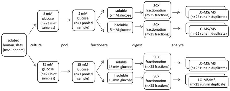
Figure 1.
Schematic showing the approach used to characterize the islet proteome under normal and high glucose conditions. Isolated human islets (handpicked from 21 pancreatic donors) were divided in half (by donor) and incubated for 24 h with either 5 mM or 15 mM glucose. Individual samples were pooled based on glucose concentration, cells were lysed, and proteins were separated based on solubility. Each of the resulting four samples was digested, and peptides were fractionated by strong cation exchange chromatography (SCX). All SCX fractions were analyzed in duplicate by reversed-phase LC-MS/MS (n = 217).

