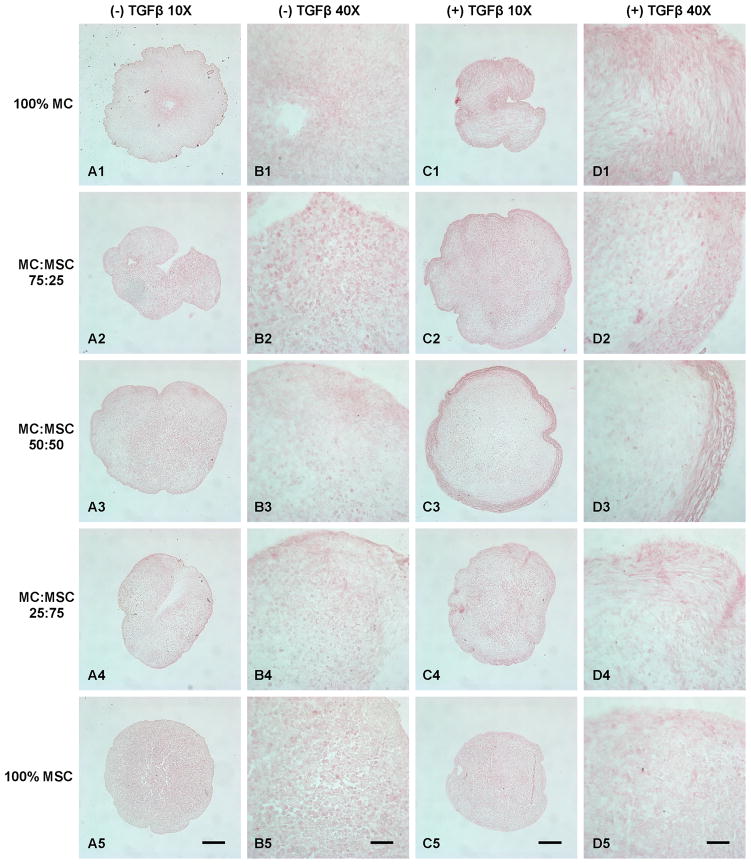
Figure 6.
Collagen type II immunohistochemistry staining of cell pellets treated with and without TGFβ. A: Cell pellets treated without TGFβ (10X). B: Cell pellets treated without TGFβ (40X). C: Cell pellets treated with TGFβ (10X). D: Cell pellets treated with TGFβ (40X). Scale bars: A, C = 200 μm; B, D = 50 μm.

