
Figure 1. The Hand1 sympathetic neuron-specific enhancer drives reporter gene expression in neurons innervating the heart.
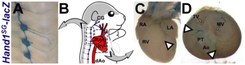
A) X-gal stained sympathetic ganglia in P0 Hand1SG-lacZ(+) mice. B) An illustration demonstrates how the CNS (gray) and the heart (red) are linked by the Hand1SG-lacZ–expressing sympathetic neurons (blue). Boxes highlight the stained tissues shown in A, C, and D. C, D) X-gal stained sympathetic nerves in P0 Hand1SG-lacZ(+) mice. In D, the atria and great vessels have been removed to provide an unobstructed view of the nerves that project from between the atrioventricular canal and outflow tract (arrowheads). Ao – aorta, CG – cervical ganglia, dAo – descending aorta, LA – left atrium, MV – mitral valve, PT – pulmonary trunk, RA – right atrium, RV – right ventricle, TV – tricuspid valve.