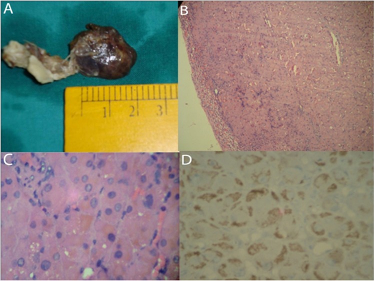
Figure 3.
(A) Macroscopic appearance of the right adrenal gland showing a black coloured circumscribed lesion 2.5 cm in maximum dimension. (B) Low power view of the tumour showing a well-circumscribed tumour with thinned out adrenal cortex in the periphery (arrow) (H&E x100). (C) High power view of the cells demonstrating the fine brown coloured pigment in the cytoplasm of individual cells (H&E x400). (D) Cytoplasmic HMB 45 positivity in the compact cells (IHC- HMB x400).

