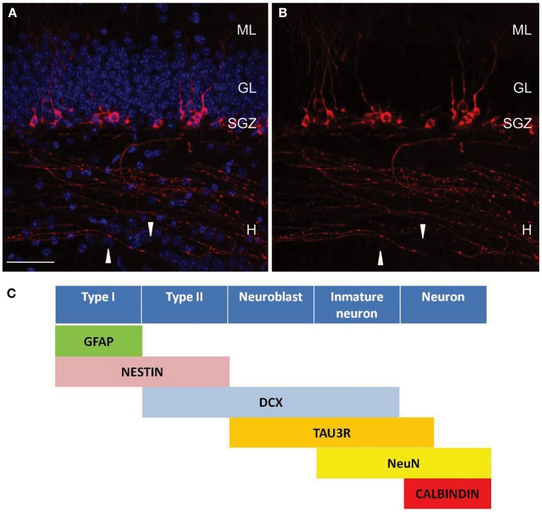
Figure 1.
Tau-3R expressing cells in the DG. (A,B) Tau-3R (red) immunohistochemistry in 2-month-old wild-type (C57BL/6) mice showing the abundance of tau-3R expressing cells along the SGZ. White arrows indicate axonal processes. DAPI staining in blue. Scale bar: 50 μm. H, hilus; GL, granular layer; SGZ, subgranular layer. (B) Shows tau-3R immunolabeling. Tau-3R antibody labeled the somatic compartment of a subpopulation of cells in the SGZ of the hippocampal DG as well as axonal processes in the hilar region and hippocampal CA3 subfield. (C) Diagram indicating the lineage and marker expression during adult neurogenesis in SGZ including tau-3R as a new marker for axonal processes [(A,B) reprinted from Journal Alzheimer Disease (Llorens-Martin et al., 2011) with permission from IOS Press].

