
Figure 2. Tissue images of masses that developed in the rasH2 mice.
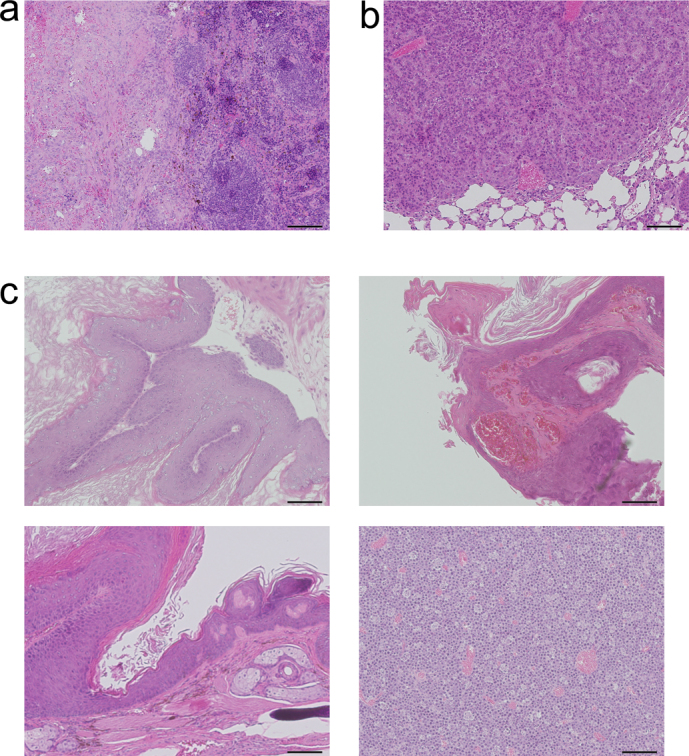
(a) A mass was observed in the spleen of 1 mouse from the CNT group, but it was found to be an inflammatory pseudotumor and not a neoplasm. (b) One mouse from the carbon black group was alive at the 26th week and was found to have developed a neoplasm in the lung, which was diagnosed as an adenoma. (c) All 10 mice in the MNU group developed forestomach tumors, with abnormal growth of squamous cells (left upper panel). Papillomas of skin were observed in 6 mice (right upper panel). Perineal tumors were found to have developed in 5 mice (left lower panel), while 2 mice developed malignant lymphoma (right lower panel). Hematoxylin-eosin staining. Scale bar: 100 μm.