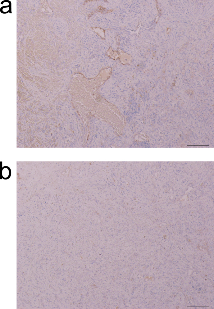
Figure 3. Immunostaining of the spleen tissue for Factor VIII.

(a) Hemangioma was diagnosed in the spleen of a mouse from the carbon black group. Immunostaining revealed vascular endothelial-like cells stained in dark brown. Atypical nucleus was not observed. (b) Inflammatory pseudotumor was diagnosed in the spleen of a mouse from the CNT group. Few cells were stained. Scale bar: 100 mm.
