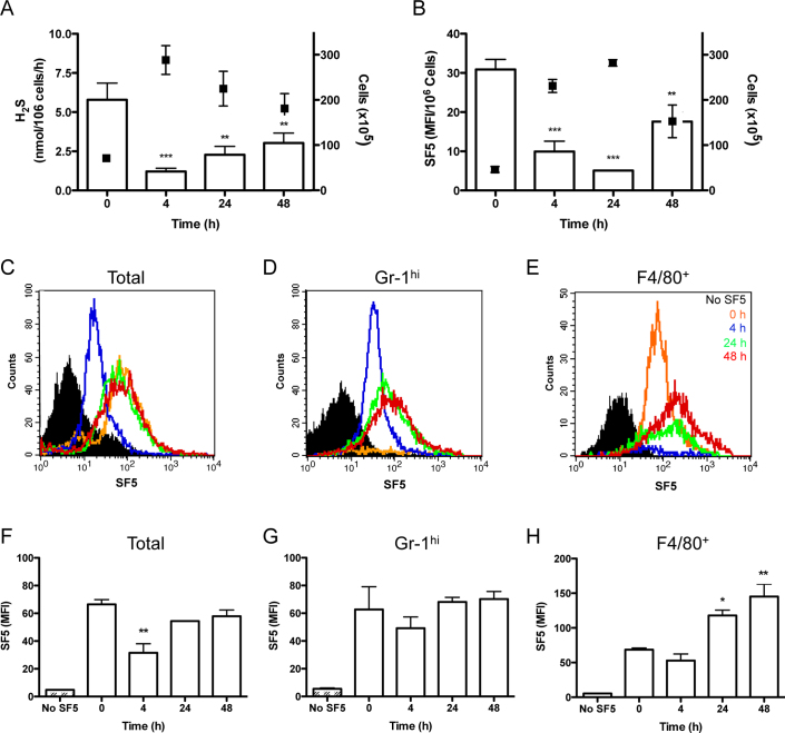
Figure 4. Mononuclear cells are an important source of hydrogen sulfide during inflammation: H2S as a mediator of resolution.
The profile of H2S synthesis by infiltrating leukocytes was compared during zymosan-induced peritonitis measured by methylene blue formation following zinc acetate capture (A) white bars; left Y-axis (n = 10) or SF5 probe (B) injected i.p. (100 μl; 10 μM) 30 min prior to euthanasia. SF5 fluorescence intensity was assessed by spectrofluorometry in peritoneal lavage is expressed as a ratio of cell numbers (n>4). Total cell numbers are overlaid (black points; right Y-axis). ***P<0.001, **P<0.01 compared by one-way ANOVA and Dunnett's post-test to zymosan alone (n>4). SF5 fluorescence within infiltrating leukocytes was analyzed by flow cytometry and represented as histograms over the time course for total infiltrating leukocytes (C) GR-1hi leukocytes (D) and F4/80+ leukocytes (E). Black filled histograms represents isotype control (C) GR-1 only (D) and F4/80 only (E). Time points are represented as 0 h; orange, 4 h; blue, 24 h; green, and 48 h; red. SF5 mean fluorescence intensity was quantified for total infiltrating leukocytes (F), GR-1hi cells (G) and F4/80+ cells (H). **P<0.01, *P<0.05 compared by one-way ANOVA and Dunnett's post-test to 0 h time point (n>4; representative of 2 experiments).

