
Figure 1. Transcription and replication of pericentromeric heterochromatin in fission yeast.
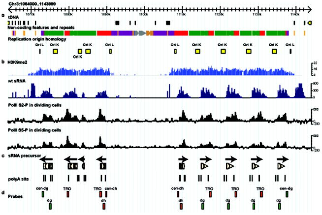
a. Pericentromeric heterochromatin on Centromere 3. dh (red), dg (green) and imr (magenta) repeats are shown, bordered by tRNA genes (brown). Replication origins (yellow) are found in each repeat. b. Tiling microarrays of K9me2 ChIP (light blue) and clusters of small RNA sequences (dark blue) from wild-type cells. ChIP-seq reads corresponding to poised (S5-P) and elongating (S2-P) RNA polymerase II enriched in dcr1Δ cells relative to WT cells are in black. c. cDNA clones (beige) from dcr1Δ cells. PolyA sites are indicated as vertical lines and correspond to peaks of PolII. Arrows indicate the direction of “Forward” transcription. d. Alignment of probes used in previous studies indicates that regions enriched for PolII11 (cen-dg) and transcriptional run-on probes1 (TRO) lie downstream of forward orientation polyA sites