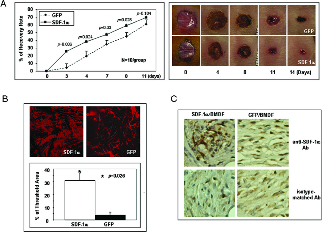
Figure 1.
SDF-1α–engineered BMDFs promote neovascularization and diabetic wound healing. (A) Left: wound healing rate expressed as percent recovery. Two groups of NOD mice were wounded and treated with mSDF-1α/BMDFs versus GFP/BMDFs. The fraction of initial wound area was measured daily by digital photography and ImageJ analysis until wounds were healed. Diabetic mice treated with mSDF-1α/BMDFs had significantly improved wound closure rates from day 3 when compared with GFP/BMDFs treated controls. Right: representative wounds at different days are shown for each group. (B) Wound blood vessel perfusion with Dil dye. Upper: representative images of Dil-stained wound blood vessels measured by laser scanning confocal microscopy at day 5 are shown for each group. Lower: Quantification of vessel density in the wounds. Percentage of threshold area covers all vessels detected as a percent of the entire wound area. mSDF-1α/BMDFs treated wounds had significantly higher vessel density compared to the control. Data are presented as mean ± SD of four wounds in each group. (C) IHC of wound tissues injected with mSDF-1α/BMDFs versus GFP/BMDFs for the detection of mSDF-1α. Stronger mSDF-1α expression (brown) was observed in mSDF-1α/BMDFs-treated wounds compared to the GFP/BMDFs treated wounds (40X).

