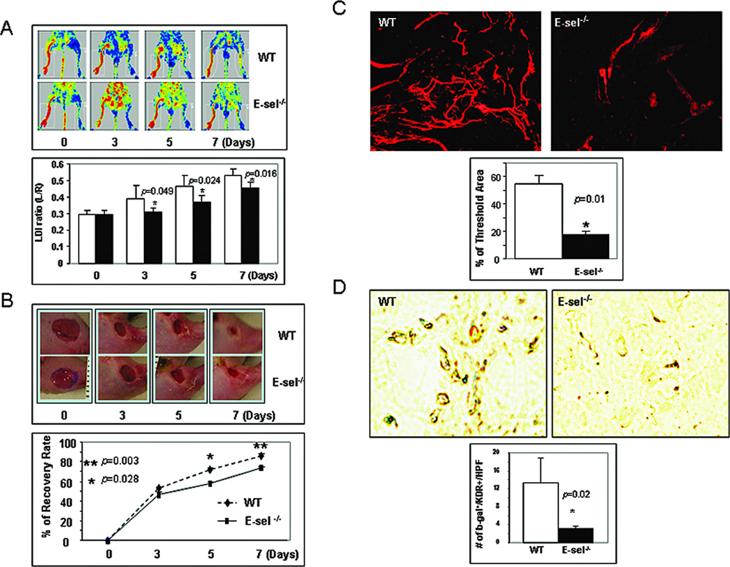
Figure 6.
Involvement of E-selectin in SDF-1α-induced EPC homing, neovascularization and wound healing in murine model of hindlimb ischemia plus cutaneous wounding. (A)Upper: representative images of non-invasive LDI measurements showing spontaneous restoration of blood flow into ischemic hindlimbs after femoral artery ligation/excision in E-sel−/− versus WT mice. Lower: Quantitative data of LDI measurements. Ratio of ischemic versus normal hindlimb between two groups of mice at various time points. Data are presented as mean ± SD from each group (n=6/group). (B) Wound closure rates in E-sel −/− versus WT. Upper: representative images of wound healing in E-sel−/− and WT mice. Deletion of E-selectin delayed wound healing. Lower: Quantitative wound closure rates in E-sel−/− versus WT mice. Data are presented as percentage wound closure (recovery), mean ± SD from each group (n=6/group). (C) Wound blood vessel perfusion with Dil dye. Upper: representative images of Dil-stained wound blood vessels detected by confocal laser scanning photography at day 7 are shown for each group. Lower: Software-assisted quantification of vessel density in the entire area of residual wounds at day 7, as percent fluorescence. Wounds from WT mice had significantly higher vessel density compared to that from E-sel−/− mice. Data are presented as mean ± SD of three wounds in each group. (D)E-sel is essential for EPC homing to the wound lesion. 1 × 10⁷ of bone marrow cells from ROSA26 (LacZ+) mice were transplanted to E-sel−/− and WT mice, respectively. Ischemic hindlimb wounds were created and SDF-1α was injected into wounds. 7 days after cell transplantation, wounds were harvested and frozen samples were subjected for X-gal (blue) and anti-KDR (brown) staining. Upper: representative images of double staining. Lower: number of double positive cells was counted from 5 randomly selected fields in wound samples. Data are percentage of mean ± SD from each group (n=3 wounds/group).

