Fig. 10.
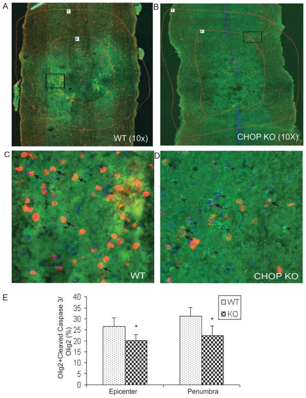
CHOP null mice exhibit less apoptotic cells after spinal cord injury. Nine 20 μm longitudinal sections (every fifth section through each cord) were immunostained and imaged under identical conditions. Representative images of (A) WT and (B) CHOP null mice (n = 4/group) were counted for colocalized Olig2+cleaved caspase 3 (yellow) and Olig2 (green) cells at the injury epicenter and penumbral region (shown with red lines 1 and 2). Adjacent GFAP-stained sections were used to define the injury epicenter. Zoomed images of the square boxes in A and B show the colocalized cells at higher magnification in (C) WT and (D) CHOP null mice. Arrowheads were used for Olig2 cells, and arrows were used for colocalized Olig2 + cleaved caspase 3 cells. (E) Quantification of Olig 2 positive cells colocalized with cleaved caspase 3 exhibited a significant 24.2% (P = 0.034) and 27.6% (P = 0.028) decrease in apoptotic cells at the injury and penumbra region, respectively, in CHOP null mice after moderate contusion injury.

