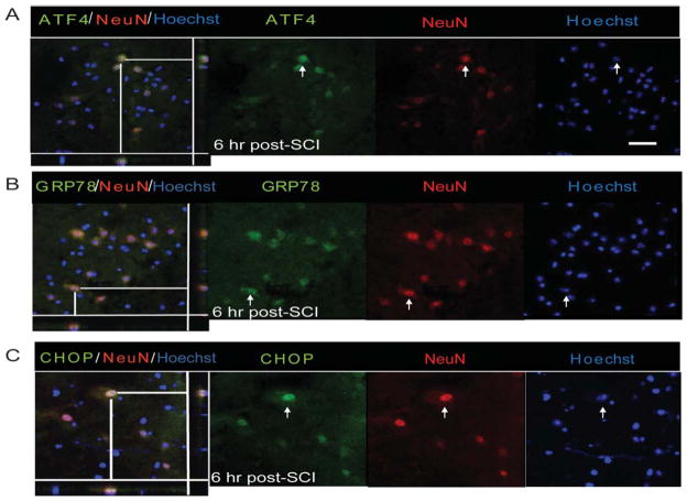
Fig. 3.
Neuronal expression of UPR markers in WT animals after SCI. Immunohistochemical analyses of longitudinal sections reveal colocalization of ATF4 (A), GRP78 (B), and CHOP (C) with NeuN, a neuron-specific marker, at the injury epicenter 6-h post-SCI. Arrows indicate the individual co-localized cells that are identified in the XZ and YZ planes of the merged images. Magnification bar = 50 μm. Identical data were observed in four independent experiments.

