Figure 2.

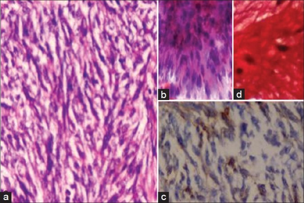
Fibrosarcoma (a) High cellularity, malignant cells forming herring bone pattern (Hist., H and E, ×400) (b) cellular smear, fascicles of malignant spindle cells (FNAC, H and E, ×400) (c) Vim + (d) VG +

Fibrosarcoma (a) High cellularity, malignant cells forming herring bone pattern (Hist., H and E, ×400) (b) cellular smear, fascicles of malignant spindle cells (FNAC, H and E, ×400) (c) Vim + (d) VG +