Figure 3.

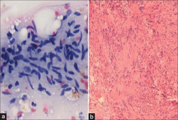
Schwannoma (a) Loosely arranged spindled vesicular nuclei with pointed ends, fibrillary stroma (FNAC, H and E, ×400) (b) Palisading spindle nuclei, verocay bodies (Histo., H and E, ×200)

Schwannoma (a) Loosely arranged spindled vesicular nuclei with pointed ends, fibrillary stroma (FNAC, H and E, ×400) (b) Palisading spindle nuclei, verocay bodies (Histo., H and E, ×200)