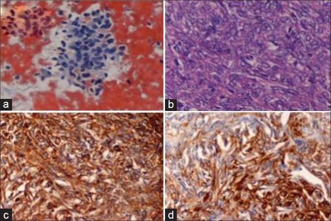
Figure 4.

Synovial sarcoma (a) Cellular smear, round to spindle nuclei, hemorrhagic background (FNAC, H and E, ×400) (b) Slit like vascular spaces surrounded by cells with thick nuclear membrane and high mitotic rate (Histo., H and E, ×400) (c) Vim + (d) CD 34 +
