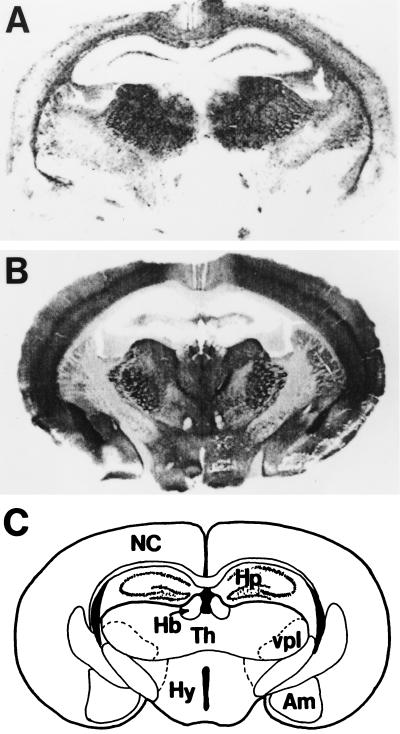
Figure 9.
Regional distribution of PrPSc deposition in Tg(MHu2M)Prnp0/0 mice inoculated with prions from humans who died of inherited prion diseases (Table 5). Histoblot of PrPSc deposition in a coronal section a Tg(MHu2M)Prnp0/0 mouse through the hippocampus and thalamus (27). (A) The Tg mouse was inoculated with brain extract prepared from a patient who died of FFI. (B) The Tg mouse was inoculated with extract from a patient with fCJD(E200K). Cryostat sections were mounted on nitrocellulose and treated with proteinase K to eliminate PrPC (209). To enhance the antigenicity of PrPSc, the histoblots were exposed to 3 M guanidinium isothiocyanate before immunostaining using anti-PrP 3F4 mAb (174). (C) Labeled diagram of a coronal sections of the hippocampus/thalamus region. NC, neocortex; Hp, hippocampus; Hb, habenula; Th, thalamus; vpl, ventral posterior lateral thalamic nucleus; Hy, hypothalamus; Am, amygdala. Photomicrographs were prepared by Stephen J. DeArmond.

