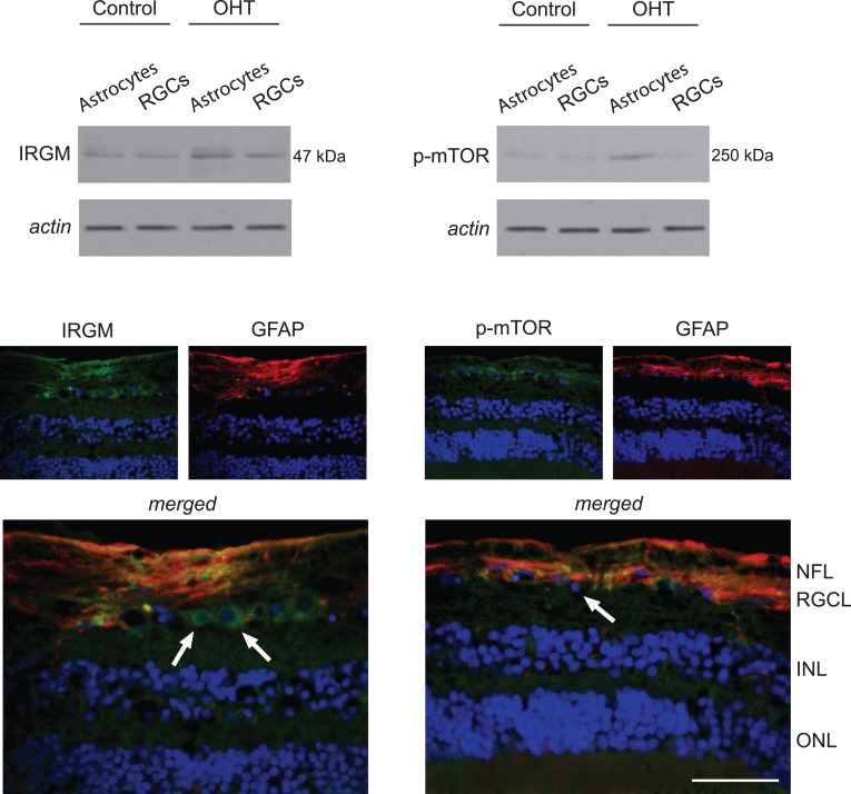
Figure 4. .
Regulation of autophagy signaling in ocular hypertensive astrocytes. Western blot analysis detected prominent up-regulation of IRGM in OHT astrocytes. In addition, probing with a phosphorylation site-specific antibody to mTOR indicated increased immunoreactivity in these samples of OHT astrocytes. Based on immunofluorescence labeling, GFAP-positive (red) astrocytes and also some GFAP-negative cells in the RGC layer, likely corresponding to RGCs (arrows), exhibited IRGM immunolabeling (green) in the OHT rat retina. However, phospho-mTOR (green) immunolabeling was detectable predominantly in GFAP-positive astrocytes. Blue corresponds to nuclear DAPI labeling. Data represent three independent sets of analyses with different samples. Scale bar 100 μm.

