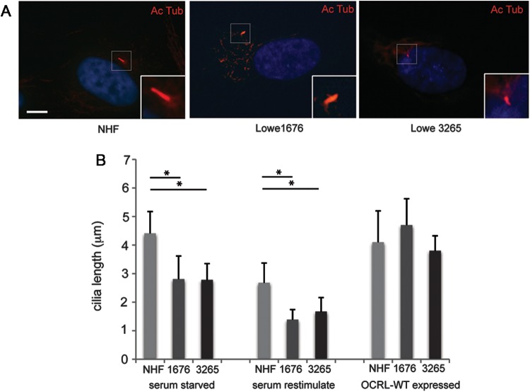
Figure 4.
Primary cilia instability in Lowe fibroblasts. (A) Representative images of NHF, Lowe 1676 and Lowe 3265 cells that were serum starved for 24 h, re-stimulated with serum for 4 h and then stained by immunofluorescence using mouse anti-acetylated α-tubulin antibody (red). Scale bar 5 μm. (B) NHF, Lowe 1676 and Lowe 3265 cells were either serum starved for 24 h or serum starved for 24 h and then re-stimulated with serum for 4 h, or transduced with OCRL WT containing lentivirus and serum starved for 24 h; different treatment groups are shown in graph separately. Immunofluorescence was performed using mouse anti-acetylated α-tubulin antibody and DAPI (blue). The cilia length was measured by two different observers (*P < 0.01, n = 3, total cells counted >100 per group, ANOVA analysis).

