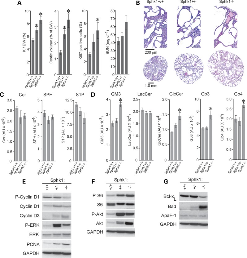
Figure 3.
The loss of Sphk1 activity exacerbates cystogenesis in jck mice. (A) PKD progression in 50-day-old Sphk1:jck males. Kidney/body weight ratio (K/BW), cyst volume, percentage of Ki67-positive cells and BUN are shown. Data shown are the mean ± SEM of 13–25 animals per group for all but percentage of Ki67-positive cells, which is 3 representative animals per group. (B) Representative H&E-stained sections from (Sphk1+/+), heterozygous mutant (Sphk1+/−) and homozygous mutant (Sphk1−/−) jck mice. (C) LC-MS analysis of bioactive lipids in kidneys of 50-day-old jck males carrying Sphk1 mutations. A gene-dosage dependent reduction in S1P levels is noted (P = 0.084 for Sphk1−/− compared with Sphk1+/+). Data shown are the mean ± SEM of three representative animals. (D) LC-MS analysis of GSL levels in kidneys of 50-day-old jck males carrying Sphk1 mutations. *P < 0.05 compared with Sphk1+/+ control. Data shown are the mean ± SEM of three representative animals. (E) Immunoblot analysis of cell-cycle regulatory protein expression in Sphk1 mutant jck mice. P-cyclin D1, phosphorylated cyclin D1; cyclin D1, total cyclin D1; cyclin D3, total cyclin D3; P-ERK, phosphorylated ERK1/2; ERK, total ERK1/2; GAPDH, glyceraldehyde-3-phosphate dehydrogenase. (F) Immunoblot analysis of the Akt-mTOR pathway in Sphk1 mutant jck mice. P-S6, phosphorylated ribosomal protein S6; S6, total ribosomal protein S6; P-Akt, phosphorylated Akt; Akt, total Akt. (G) Immunoblot analysis of proteins regulating apoptosis in Sphk1 mutant jck mice. GAPDH staining was used to control for loading differences.

