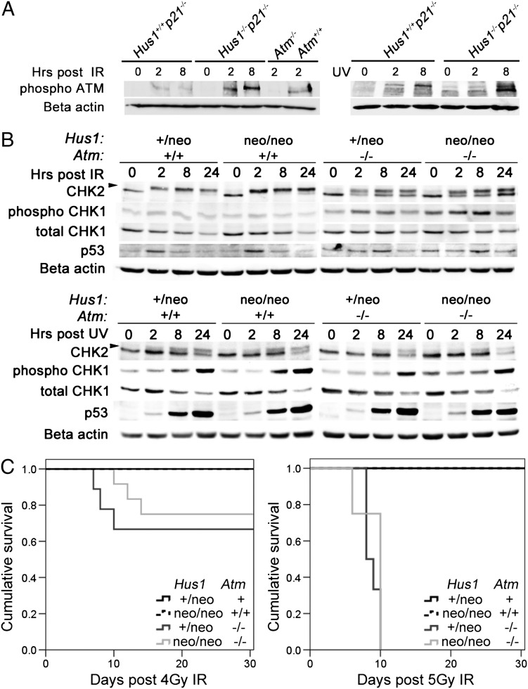
Figure 6.
DNA damage signaling in Atm/Hus1 double-mutant MEFs. (A) Immortalized MEFs of the indicated genotypes were treated with 20 Gy IR or 65 J/m2 UV, and total protein lysates were prepared at 0, 2 and 8 h post-treatment. Samples were immunoblotted using antibodies specific for phospho-Ser1981-ATM, or beta-actin as a loading control. Similar results were obtained with an independent set of Hus1−/−p21−/− and matched control Hus1+/+p21−/− MEFs (data not shown). (B) Primary MEFs at passage 1 or 2 were treated with 30 Gy IR or 65 J/m2 UV, and total cell protein lysates were prepared at 0, 2, 8 and 24 h post-treatment and immunoblotted for CHK1, phosphoSer345-CHK1, CHK2 and p53. The arrow indicates the position of phosphorylated CHK2. Beta-actin was used as a loading control. (C) Kaplan–Meier survival curves show the radiation sensitivity of mice of the indicated genotypes, following exposure to 4 Gy (left) or 5 Gy (right) IR. The cohort treated with 4 Gy included Hus1+/neoAtm+ (n=7; Atm+ is a combination of Atm+/+ and Atm+/−), Hus1neo/neoAtm+/+ (n=6), Hus1+/neoAtm−/− (n=9) and Hus1neo/neoAtm−/− (n=12) mice. The cohort treated with 5 Gy included Hus1+/neoAtm+ (n= 4), Hus1neo/neoAtm+/+ (n=6), Hus1+/neoAtm−/− (n=6) and Hus1neo/neoAtm−/− (n=4) mice. There were no significant differences in survival between Hus1+/neoAtm−/− and Hus1neo/neoAtm−/− mice after 4 Gy (P=0.295) or 5 Gy (P=0.366) as determined by the log-rank test.

