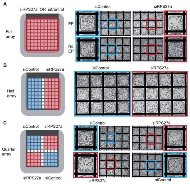
Fig. 3.
Electroporation of functional siRNAs bound within microwell arrays and evaluation of cellular phenotypes. A) Left-Schematic showing that the full area microwell arrays were coated with either siRPS27a or siControl. Right-Death phenotypes were observed only in the siRPS27a-coated microwells and only under electroporation conditions. No death was observed in siRPS27a-coated microwells without electroporation. B) Left-Schematic showing half areas of the microwell array coated with siRPS27a or siControl. Right-Death phenotypes were observed only on the siRPS27a-coated half. C) Left-Schematic showing quadrant areas of the microwell array coated with siRPS27a or siControl. Right-Death phenotypes were observed only in quadrants containing siRPS27a. Experiments were performed with HeLa cells and phenotypes assessed 48 h post-electroporation.

