
Figure 3.
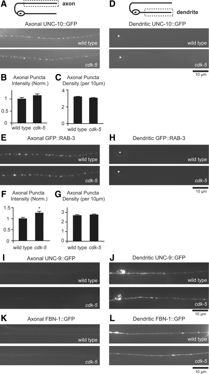
The polarized distribution of axonal and dendritic markers in DB motor neurons is not affected in cdk-5 mutants. A, Schematic diagram of a DB motor neuron (top panel). The boxed region denotes that the axon was imaged for data presented in A–C, E–G, I, and K. Representative images of UNC-10::GFP in DB axons of wild-type and cdk-5 mutant animals (bottom panels). B, C, Quantification of UNC-10::GFP puncta intensity (B) and density (C) in axons of wild-type (n = 21) and cdk-5(gm336) (n = 20) mutant animals. D, Schematic diagram of a DB motor neuron (top panel). The boxed region denotes that the dendrite was imaged for data presented in D, H, J, and L. Representative images of UNC-10::GFP in DB dendrites of wild-type and cdk-5(gm336) mutant animals (bottom panels). E, Representative images of GFP::RAB-3 in DB axons of wild-type and cdk-5(gm336) mutant animals. F, G, Quantification of GFP::RAB-3 puncta intensity (F) and density (G) in axons of wild-type (n = 22) and cdk-5(gm336) (n = 21) mutant animals. H, Representative images of GFP::RAB-3 in DB dendrites of wild-type and cdk-5(gm336) mutant animals. Values that differ significantly from wild-type (Student's t test) are denoted on graphs (*p ≤ 0.01). I, Representative images of UNC-9::GFP in DB axons of wild-type and cdk-5(gm336) mutant animals. J, Representative images of UNC-9::GFP in DB dendrites of wild-type and cdk-5(gm336) mutant animals. K, Representative images of FBN-1::GFP in DB axons of wild-type and cdk-5(gm336) mutant animals. L, Representative images of FBN-1::GFP in DB dendrites of wild-type and cdk-5(gm336) mutant animals.