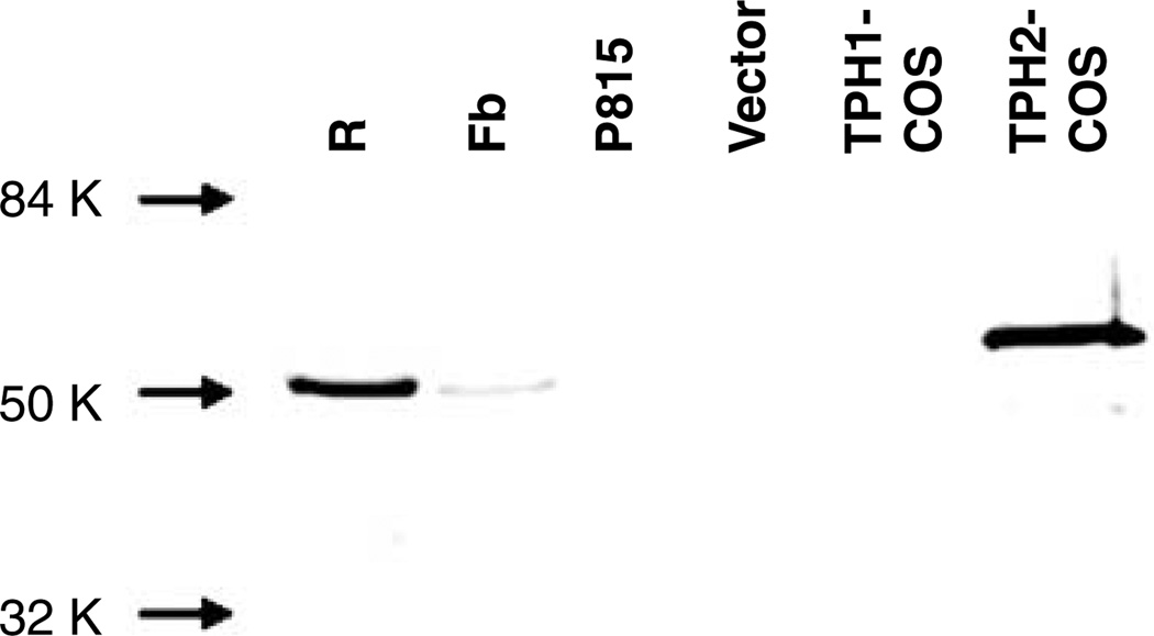
Figure 1.
Immunoblotting with TPH2-specific antiserum. Rodent brain tissue samples (40 µg), P815 whole-cell homogenate (30 µg) and COS whole-cell homogenates (1 µg) were run on sodium dodecylsulfate-polyacrylamide gel electrophoresis and transferred to nitrocellulose for immunoblotting. The TPH2-specific antiserum TPH2-6361 detected a dominant ~55 kDa and a minor ~47.5 kDa band in whole-cell homogenates from COS cells overexpressing TPH2 (TPH2-COS). The antiserum detected an ~51 kDa band in total protein from a murine midbrain slice containing raphe nuclei (R) and murine forebrain (Fb). The antiserum did not detect bands in whole-cell homogenates from pcDNA 3.1-transfected COS cells (Vector), COS cells overexpressing TPH1 (TPH1-COS) or in P815 cells, a murine mastocytoma cell line that expresses TPH1 exclusively. TPH, tryptophan hydroxylase.

