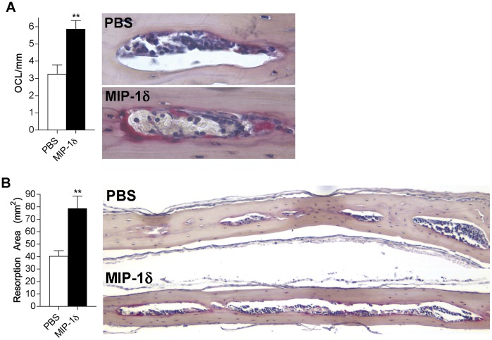
Figure 4. MIP-1δ promotes osteoclastogenesis and bone resorption in vivo.
Mice (5/group) were administered MIP-1δ (1 ng/ml) or PBS subcutaneously over the calvariae twice each day for 5 days. Calvariae were removed and paraffin-embedded sections were generated for histomorphometric analysis. (A) OCL were identified by TRAP staining, counted under light microscopy (right, 400X), and reported relative to mm of marrow cavity surface (left). (B) Resorption area (left) was determined by measuring the total area occupied by marrow cavities within each calvarial section (right, 100X). Data are representative of two independent experiments, expressed as the mean (n = 10) ± s.e.m, and were analyzed by unpaired, two-tailed Student’s t-test. **, p<0.01.

