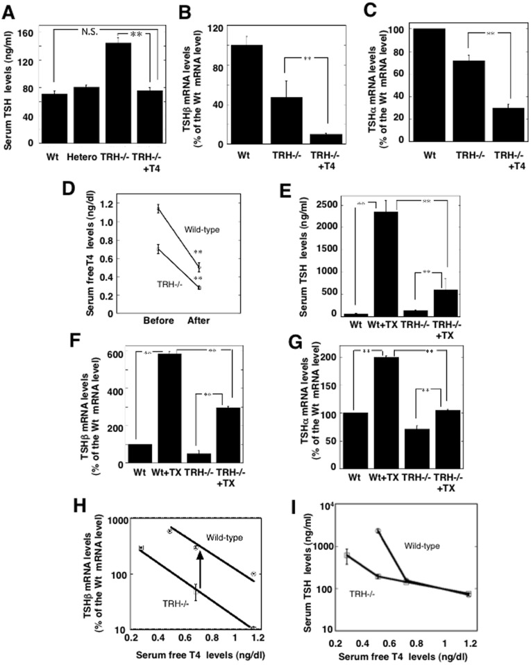
Figure 1. The hypothalamic-pituitary-thyroid axis in TRH deficient mice.
A) Serum TSH levels were measured in wild-type mice (Wt), TRH knockout mice (TRHKO) heterozygotes (Hetero) and homozygotes (TRH−/−) and TRHKO supplemented with thyroid hormone (TRH−/− + T4). TRHKO (n = 7) were supplemented with thyroid hormone by the daily injection of T4 (1.5 μg/100 g body weight) for 14 days to be euthyroid (n = 6). The serum TSH level in TRH−/−+T4 mice was similar to that in the wild-type (n = 7) and heterozygous mice (n = 6). Values are represented as the mean ± SEM. **, p<0.01; *, N.S., not significant. B) TSHβ mRNA levels were measured in pituitaries of the wild-type mice, TRHKO, and TRHKO supplemented with thyroid hormone (TRH−/−+T4). The TSHβ mRNA level in TRH−/− was decreased to about 50% of that in the wild-type, and that of TRH−/−+T4 mice was further decreased to about 10% (n = 3, p<0.01). **, p<0.01. C) TSHα mRNA levels showed a similar profile to the TSHβ mRNA level shown in Fig 1B. However, the change in TSHβ mRNA was more profound than that in TSHα mRNA. **, p<0.01. D) Two weeks after thyroidectomy, serum free T4 levels were significantly reduced in both the wild-type mice (n = 6) and TRHKO mice (TRH−/−) (n = 5). The serum T4 levels were significantly lower in TRH−/− mice than wild-type mice. **, p<0.01. E) Reflecting hypothyroidism induced by thyroidectomy (TX), the serum TSH level in the wild-type mice showed an approximately 25-fold increase (Wt+TX, n = 6), but TRHKO mice (TRH−/−) showed only about a 6-fold increase (TRH−/−+TX, n = 6). **, p<0.01. F) The TSHβ mRNA levels in thyroidectomized wild-type mice (Wt+TX) were significantly increased after two weeks to approximately 6-fold the control value (Wt) (n = 3). However, the TRHKO mice (TRH−/−) showed a lesser increase (TRH−/−+TX)(n = 3). **, p<0.01; G) A similar profile was observed in TSHα mRNA levels in the pituitary. But a milder effect was observed compared to those of TSHβ mRNA levels. **, p<0.01; n = 3. H) Correlation between serum thyroid hormone levels and the corresponding pituitary TSHβ mRNA levels. Circles represent values for the wild-types and the squares, those for TRH knockout mice (TRH−/−). When comparing the slopes of the curve, it was demonstrated that TRH altered the basal activity and responsiveness of the TSHβ gene to thyroid hormone. The arrow indicates the effect of TRH, taking approximately 80% responsibility for the value of the TSHβ mRNA level in the wild-type pituitary. I) Correlation between serum thyroid hormone levels and the corresponding serum TSH levels. In mild hypothyroidism, the serum TSH levels in TRHKO mice (TRH−/−) were similar to those in the wild-type mice. However a lack of TRH induced a marked impairment of the serum TSH level in severe hypothyroid status.

