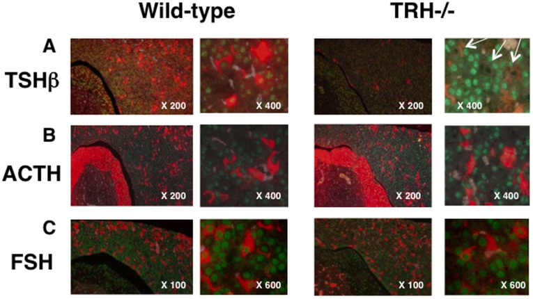
Figure 3. Expression and regulation of NR4A1 by TRH in pituitary thyrotrophs in vivo.
The expression of NR4A1 in pituitary thyrotrophs was confirmed by double fluorescent immunohistochemistry. In numbers of cells in the anterior pituitary NR4A1 were expressed and stained as green signals in the nucleus. A) Numbers of cells expressing NR4A1 were also stained with antibody against TSHβ (red signals) in the wild-type pituitary (left panel). As shown in the right panel, the intensity and number of the TSHβ-immunopositive cells were remarkably decreased in the TRH-deficient pituitary, and at X400, most of the NR4A1 expression was reduced or lost in these cells as seen as black dots (indicated by white arrows). B) As observed in Fig 4B and C, the expression of ACTH (B) and FSH (C) was also observed as red signals in the cytoplasm in the anterior lobe. As expected, these ACTH- and FSH- positive cells expressed NR4A1, but the staining intensity was not altered in the TRH-deficient pituitary (right panel).

