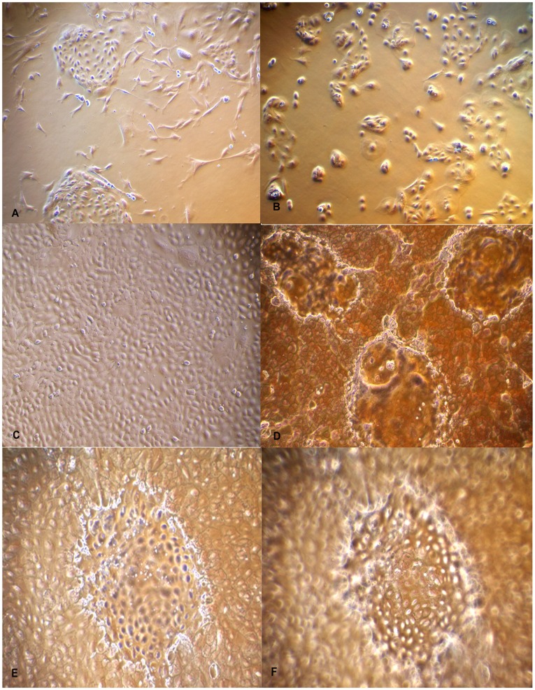
Figure 1. Photomicrographs of isolation and culture of Buffalo Mammary Epithelial cells (BuMECs).
A: Mixed population of epithelial and fibroblast cells (×100); B: Purified BuMEC seeded at low density forming islands (×100); C: Confluent mono layer of BuMECs showing cobble stone morphology (×100); D: Post confluent stage BuMECs forming dome structure (×100); E: Phase contrast image of dome structure with focus on the monolayer (×100); F: Phase contrast image of dome structure with focus set at the top of dome (×100). The dome structure represents a raised layer of cells above the plastic substratum.

