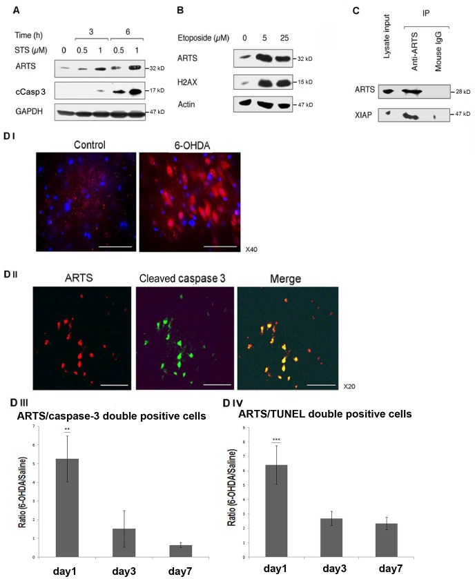
Figure 1. Up-regulation of ARTS levels is associated with induction of apoptosis in SH-SY5Y cells and in 6-OHDA treated rat brains.
A. Western blot (WB) analysis shows a strong increase in the expression levels of ARTS upon treatment of SH-SY5Y cells with the apoptotic inducer staurosporine (STS). This elevation in levels of ARTS is associated with an increase in caspase-3 activation. GAPDH was used as a loading control. B. A significant increase in levels of ARTS is seen in response to treatment with etoposide inducing apoptosis in SH-SY5Y cells. This strong up-regulation of ARTS is associated with a corresponding increase in expression of the apoptotic marker H2AX. Actin was used as a loading control. C. Endogenous ARTS binds to XIAP in SH-SY5Y cells. Immunoprecipitation assay (IP) with monoclonal anti ARTS antibody was performed on lysates from SH-SY5Y cells. Mouse IgG served as control for co-precipitation of the specific antigen. DI. A representative picture (viewed with x40 objective) showing immunofluorescence (IF) staining using anti-ARTS antibody in Substantia nigra pars compacta (SNpc) of rat brain treated with 6-hydroxy dopamine (6-OHDA) as compared to control injected with saline. 6-OHDA was injected into the left medial forebrain bundle of these rats, from where it is transported to the SN. A significant increase in expression of ARTS is shown in these cells (red). DAPI staining of nuclei is shown in blue. Scale bar represents 50 µm. DII. Co-localization of ARTS (red) and cleaved caspase-3 (green) is seen in a representative rat brain section of 6-OHDA treated rat viewed using x20 objective. Scale bar represents 100 µm. DIII, DIV. Percent of ARTS/cleaved caspase-3 and ARTS/TUNEL double positive cells in SNpc of 6-OHDA treated rats compared to saline injected controls. These pictures demonstrate the levels of ARTS among apoptotic neurons. Counts were done in sections from brains after one, three and seven days following injection of 6-OHDA or saline. Figures present mean ± SEM of the ratio between the percent of double positive cells in 6-OHDA brains relative to the average percent of double positive cells in saline treated brains, at each time point. A significant increase in percentage of ARTS/caspase-3 and ARTS/TUNEL double positive SN neurons is seen 24 hours after injection of 6-OHDA as compared to controls (significance levels were calculated from data shown in Table S1; **P<0.01, ***P<0.001 for difference from same day control by ANOVA with Bonferroni post hoc test). This suggests that ARTS may play an important role in promoting susceptibility to 6-OHDA-induced apoptosis in SN neurons.

