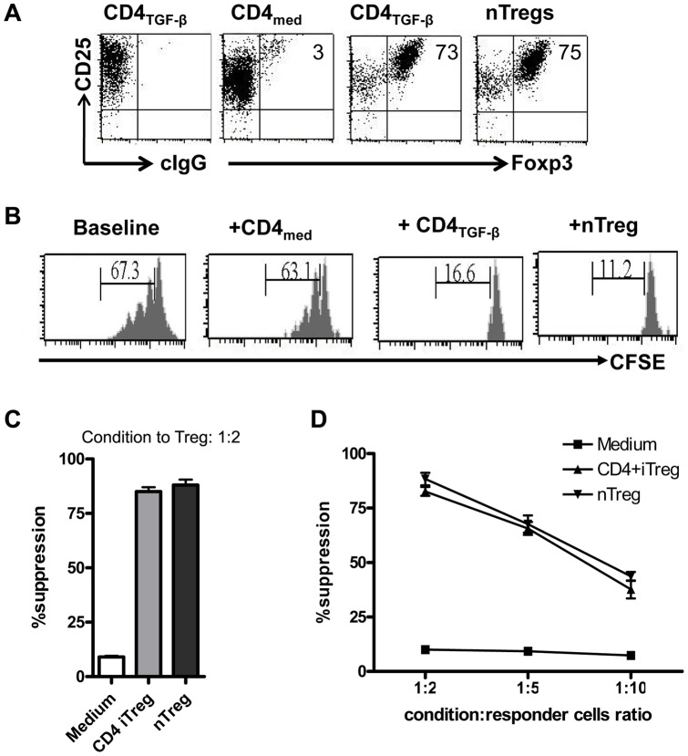
Figure 1. In vitro induction of regulatory T (iTreg) cells by TGF-β.
Naive CD4+CD25− cells were stimulated with anti-CD3/CD28 coated beads with IL-2 in the presence (CD4TGF-β) and absence (CD4med) of TGF-β for 5–6 days. nTreg cells were splenic CD4+CD25+ cells that were sorted and expanded with anti-CD3/CD28 coated beads with IL-2 for 6–7 days. (A). FoxP3 expression was determined by flow cytometry with anti-Foxp3 antibody. cIgG, control IgG. (B). T cells labeling with CFSE were stimulated with anti-CD3 with or without CD4 condition cells (ratio of CD4 condition to T responder = 1∶2) for three days and CFSE dilution was identified on the CD4+ cell gate. (C). T cell proliferation was determined by 3H-thymidine incorporation assay. (D). The T cell proliferation was determined in the different ratios of CD4 conditioned cells and T responder cells. Data was representative or mean ± SEM of three independent experiments.

