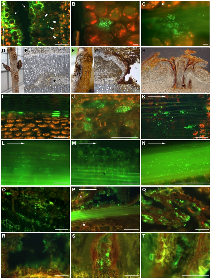
Figure 2. The infection process of P. syringae pv. aesculi and bacterial behaviour in planta.
The infection process up to 18 weeks after inoculation visualized by immunocytochemically labelling of P. syringae pv. aesculi. Arrows in A, C, K, L, M, N and P indicate the longitudinal direction. Scale bars indicate 100 µm in all micrographs except B and C where 10 µm is indicated. (A) Six days after inoculation, when the lining of lignified and suberized cells is prominent, clusters of bacteria are observed in deeper parenchyma tissues (arrowheads) by immunolabelling. Furthermore, bacteria remain in the original inoculation cut (arrows). The image shown is taken from a longitudinal section. (B) A bacterial accumulation in a cortex parenchyma cell observed in a longitudinal section of an infection site sampled 14 days after inoculation. Besides the large cluster, several individual bacteria are scattered in intercellular spaces. (C) A longitudinal section of phloem tissue 6 days post inoculation showing the decoration of bark fibres by P. s. pv. aesculi seemingly following the vertical orientation of these elements. (D, E) Close-up and micrograph of transversal section of a mock-inoculated site on a horse chestnut plant 7 weeks after inoculation. The asterisk points to site of wounding during inoculation. (F, G, H) Close up and transversal sections of inoculation sites 15 weeks after inoculation. Note partially restored development of secondary xylem despite infection. Asterisks point to site of wounding during inoculation. (I, J) Transversal sections of cortex in mock-inoculated (I) and infected (J) horse chestnut 12 days after inoculation. Note ablated plant cells filled with bacteria in J. (K) Clusters of bacteria in a longitudinal section of the xylem close to the inoculation site, 4 weeks after inoculation. (L–N) Longitudinal sections of the cambium region with bacteria in intercellular spaces of phloem parenchyma (L), ray parenchyma (M) and along phloem fibres (N), 3 weeks after inoculation. (O) P. s. pv. aesculi in intercellular spaces observed in a transversal section of the cambial zone at 4 weeks after inoculation. (P) Bacteria decorating a bundle of phloem fibres which is surrounded by newly formed periderm (*) and degenerating parenchyma 4 weeks after inoculation. (Q) Bacteria colonizing necrotic bark tissue at 10 weeks after inoculation. (R-T) Transversal sections of enclosures surrounded by periderm with bacteria ex planta at 4, 10 and 18 weeks after inoculation, respectively.

