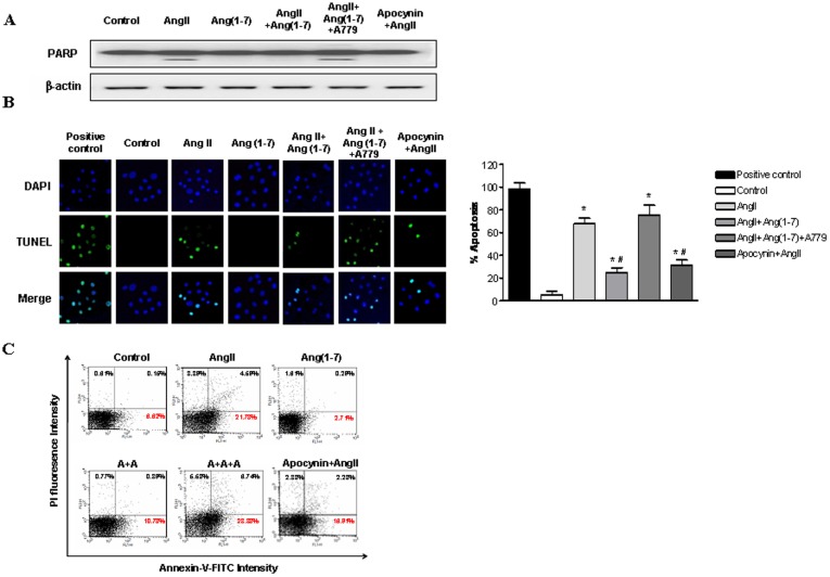
Figure 6. Effects of Ang-(1-7) on AngII-induced ROS-related apoptosis.
A, Western blotting of PARP and cleaved caspase-3 in NRK-52E cells exposed Ang II (10−6 M) with or without Ang-(1-7) (10−6 M), A779 (10−6 M), and apocynin (100 µM). B, TUNEL staining of NRK-52E cells. Apoptotic nuclei are depicted by green fluorescence. TUNEL-positive nuclei in control cells after DNase I treatment. Cells were exposed to Ang II (10−6 M) with or without Ang-(1-7) (10−6 M), A779 (10−6 M), and apocynin (100 µM). TUNEL-positive nuclei were counted from random fields per slide and expressed as the percentage of apoptotic cells (apoptotic nuclei/total nuclei ×100%). C, Flow cytometric analysis of NRK-52E cells with annexin V-FITC/PI double staining. NRK-52E cells exposed to Ang II (10−6 M) with or without Ang-(1-7) (10−6 M), A779 (10−6 M), and apocynin (100 µM). Values are the mean ± S.E.M. (n = 4). *p<0.05 versus control; #p<0.05 versus Ang II-stimulated cells.

