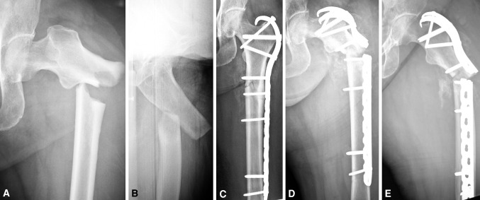
Fig. 1A–E.
(A) AP and (B) oblique radiographs show a characteristic fracture of the femur associated with bisphosphonate therapy; the lateral cortex at the fracture site is thickened, which alters the relationship between the canal and cortical surfaces. (C) An AP image is shown that was obtained after open reduction and internal fixation with a proximal femoral hook LCP Plate. (D) AP and (E) lateral radiographs obtained after hardware failure 3 months after fracture surgery show plate breakage.

