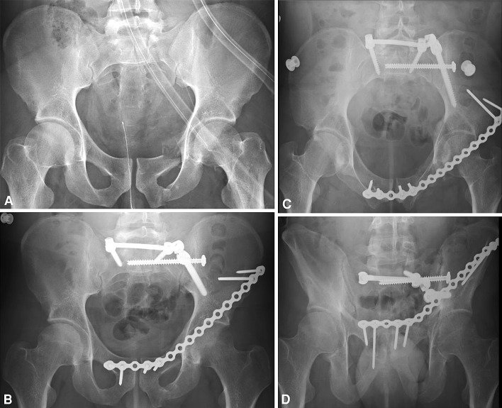
Fig. 4A–D.
(A) Preoperative AP and postoperative (B) AP, (C) inlet, and (D) outlet pelvic radiographs show posterior spinopelvic fixation and unilateral left APIF with a precontoured reconstruction plate and locking screws. The posterior ring was reduced and stabilized before anterior ring fixation.

