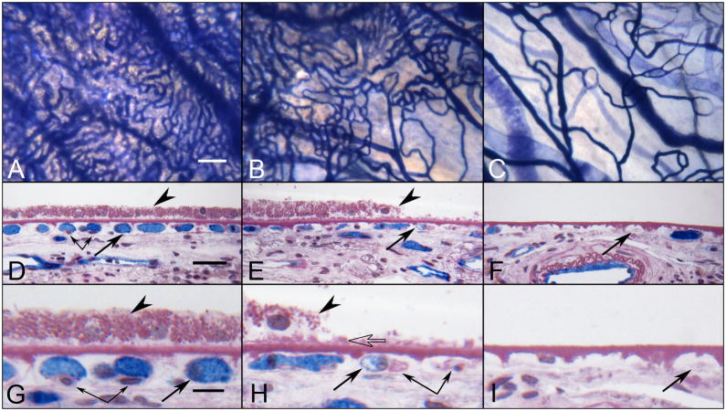
Figure 2.
Alkaline phosphatase (APase)-incubated choroid from a 88 year-old Caucasian male with GA showing the nonatrophic region (A,D&G), border region (B,E&H) and atrophic region (C,F&I) in flat view prior to embedment (A-C) and in cross sections stained with PAS and hematoxylin (D–I). Blue APase reaction product is present in viable blood vessels only and is most prominent in neovascularization. (A) APase stained capillaries in the nonatrophic region have broad diameter lumens filled with serum APase (arrow in D&G), with endothelial cells and pericytes underlying viable RPE (arrowhead in D&G). At the border region, RPE appear hypertrophic (arrowhead in E&H), capillaries appear constricted (arrow in E&H), and some have completely degenerated (paired arrows H). A thin basal laminar deposit is associated with Bruch’s membrane (open arrow). In the atrophic region, many capillaries have degenerated leaving only remnants of basement membrane material (arrows in F&I). (scale bar = 100 μm in A–C, 30 μm in D–F, 10 μm in G–I) [Figure 10 from McLeod et al, Invest. Ophthalmol. Vis. Sci 50:4982-4991, 2009 (McLeod et al., 2009) with permission]

