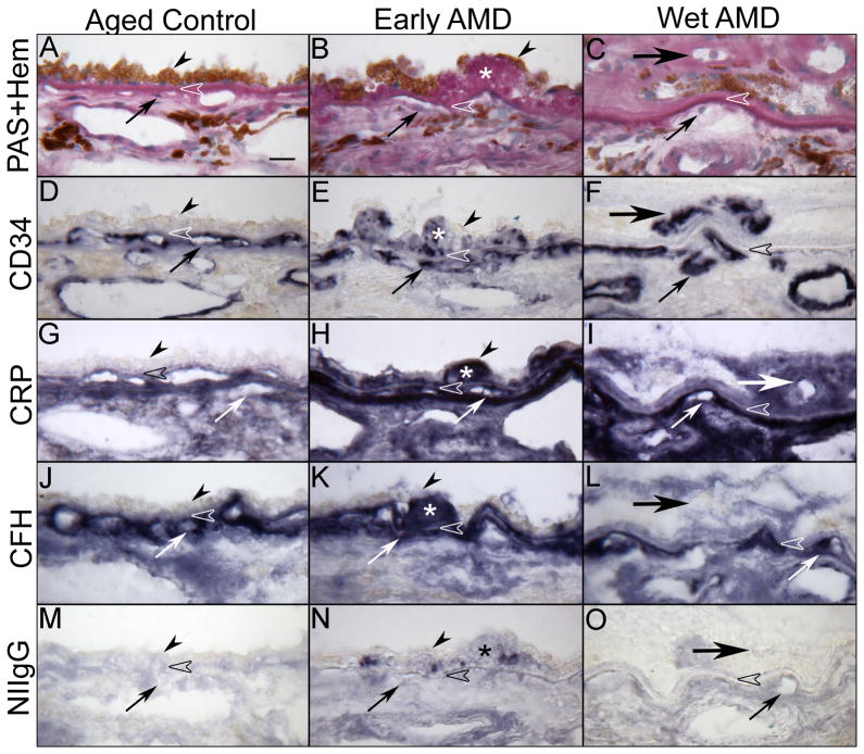
Figure 8.
Immunolocalization of C-reactive protein (CRP) and complement factor H (CFH) in submacular choroid from aged control, early and late wet AMD eyes. Periodic acid-Schiff’s (PAS) and hematoxylin (Hem) staining shows morphological features of the choroid from aged control (A), drusen (asterisk) in early AMD (B) and CNV (large arrow) anterior to RPE in wet AMD (C). Pigment in immunostained sections was bleached from RPE and choroidal melanocytes. Immunostaining of CD34 is associated with CC (small arrow) and large choroidal vessels appear morphologically normal with broad lumens in aged control (D), whereas CC lumens appear irregular and constricted in early (E) and wet AMD (F). In aged control choroid, CRP (G) and CFH (J) are prominently localized to the CC, intercapillary septa (ICS) and BrM (open arrowhead). CRP immunoreactivity is significantly increased in early (H) and late AMD (I) choroids compared to the aged control and appear more diffuse in choroidal stroma. CFH in early AMD (K) is comparable to aged control, whereas it is significantly decreased in wet AMD (L). Drusen are intensely labeled with CRP and CFH (H and K). Note that in wet AMD the CNV (large arrow), intensely labeled with CD34 antibody (F), has more CRP and less CFH (I and L). Nonimmune rabbit IgG (NIIgG) yields a very weak to negative reaction product except in drusen (M, N, O). [Figure 3 from Bhutto et al British Journal of Ophthalmology 95:1323-1330, 2011 (Bhutto et al., 2011) with permission]

