FIGURE 5.

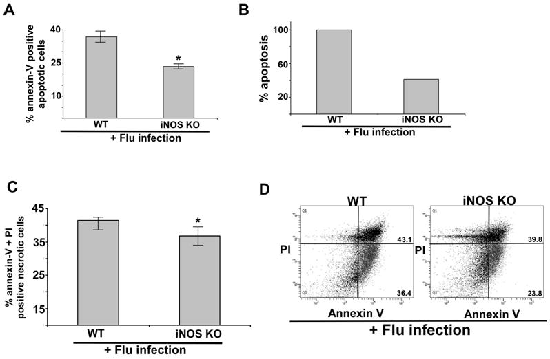
iNOS expression is required for apoptosis during flu infection of primary macrophages. (A) Percentage of annexin V positive cells (apoptotic cells) were detected by FACS analysis of flu infected (48h post-infection) wild-type (WT) and iNOS knock-out (KO) primary bone marrow derived macrophages (BMDMs). Annexin V staining quantified by FACS represents mean ± SEM from four independent experiments performed in triplicate, *p < 0.05 by two-tailed t test. (B) The values presented in Fig. 5A were utilized to calculate % apoptosis in flu infected WT and iNOS KO BMDMs. Apoptosis in flu infected WT BMDMs are denoted as 100% apoptosis. (C) Percentage of annexin V and PI positive cells (necrotic cells) were detected by FACS analysis of flu infected (48h post-infection) WT and iNOS KO) BMDMs. Annexin V + PI staining quantified by FACS represents mean ± SEM from four independent experiments performed in triplicate, *p = 0.09 by two-tailed t test. (D) A representative FACS showing percentage of apoptotic (lower right quadrant – annexin V positive and PI negative cells) and necrotic (upper right quadrant – annexin V and PI positive cells) cells following flu infection WT and iNOS BMDMs.
