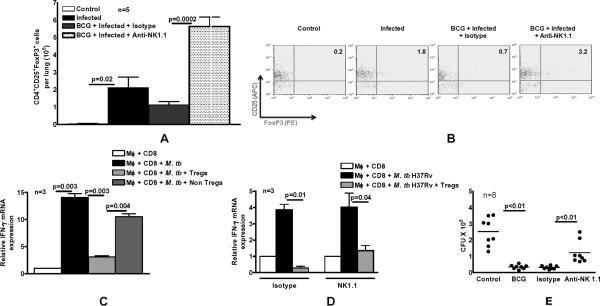
Fig. 3. NK1.1+ cells inhibit expansion of immunosuppressive Tregs after BCG vaccination and challenge with M. tb H37Rv.
C57BL/6 mice (3-8 mice per group) were unimmunized or immunized subcutaneously with 106 CFU of BCG in 100 μl of PBS. Some BCG-vaccinated mice were treated with anti-NK1.1, or isotype control Ab (0.3 mg per mouse 0, 24 and 48 h after vaccination). Sixty days after BCG vaccination, mice were challenged with 50–100 CFU of M. tb H37Rv by aerosol. A. Thirty days post-infection, CD4+CD25+FoxP3+ T-cells in the lungs were measured by flow cytometry. B. A representative flow cytometry result of lung cells is shown. We gated on lung CD4+ cells and then gated on CD25+ and FoxP3+ expressing cells. C. M. tb H37Rv-expanded CD4+CD25hi cells are immunosuppressive. Three C57BL/6 mice were immunized subcutaneously with 106 CFU of BCG. One week later, CD4+ and CD11b+ cells from spleens and peripheral lymph nodes were isolated and cultured with γ-irradiated M. tb H37Rv. After 72 h, CD4+CD25hi cells and CD4+CD25- cells were isolated and cultured in Transwells, within large wells containing CD8+ and CD11b+ cells obtained from mice one week after BCG vaccination and γ-irradiated M. tb H37Rv. After 72 h, the Transwells were removed, and IFN-γ mRNA was quantified in the cells in the large wells by real-time PCR. D. C57BL/6 mice (3 mice per group) were immunized subcutaneously with 106 CFU of BCG. Some BCG-vaccinated mice were treated with anti-NK1.1, or isotype control Ab (0.3 mg per mouse 0, 24 and 48 h after vaccination). Sixty days after BCG vaccination, mice were challenged with 50–100 CFU of M. tb H37Rv by aerosol. Thirty days post-infection, CD4+CD25hi Tregs from pooled lung, spleen and mediastian lymph nodes were isolated and cultured in Transwells, within large wells containing CD8+ and CD11b+ cells from BCG-vaccinated mice and γ-irradiated M. tb H37Rv. After 72 h, the Transwells were removed, and IFN-γ mRNA was quantified in the cells in the large wells by real-time PCR. Mean values and SEs are shown. E. C57BL/6 mice (8 mice per group) were unimmunized or immunized subcutaneously with 106 CFU of BCG. Some BCG-vaccinated mice were treated with anti-NK1.1, or isotype control Ab (0.3 mg per mouse 0, 24 and 48 h after vaccination). Sixty days after BCG vaccination, mice were challenged with 50–100 CFU of M. tb H37Rv by aerosol. Thirty days post-infection, lungs were homogenized and plated on 7H11 agar with THC, and CFU per lung were counted after 3 weeks.

