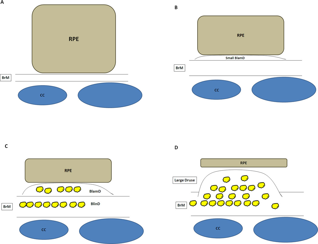
Figure 3.
Schematic diagram of degenerative changes to the RPE and Bruch’s membrane during the progression of AMD. A) Normal cuboidal morphology of the RPE. Bruch’s membrane (BrM) is normal thickness. CC, choriocapillaris. B) Flattening of the RPE with early basal laminar deposit (BlamD). C) Further flattening of the RPE with development of more advanced basal laminar deposits and development of basal linear deposits (BlinD). Heterogeneous debris (Yellow deposits) accumulates in both BlamD and BlinD. D) Atrophic RPE overlying large druse.

