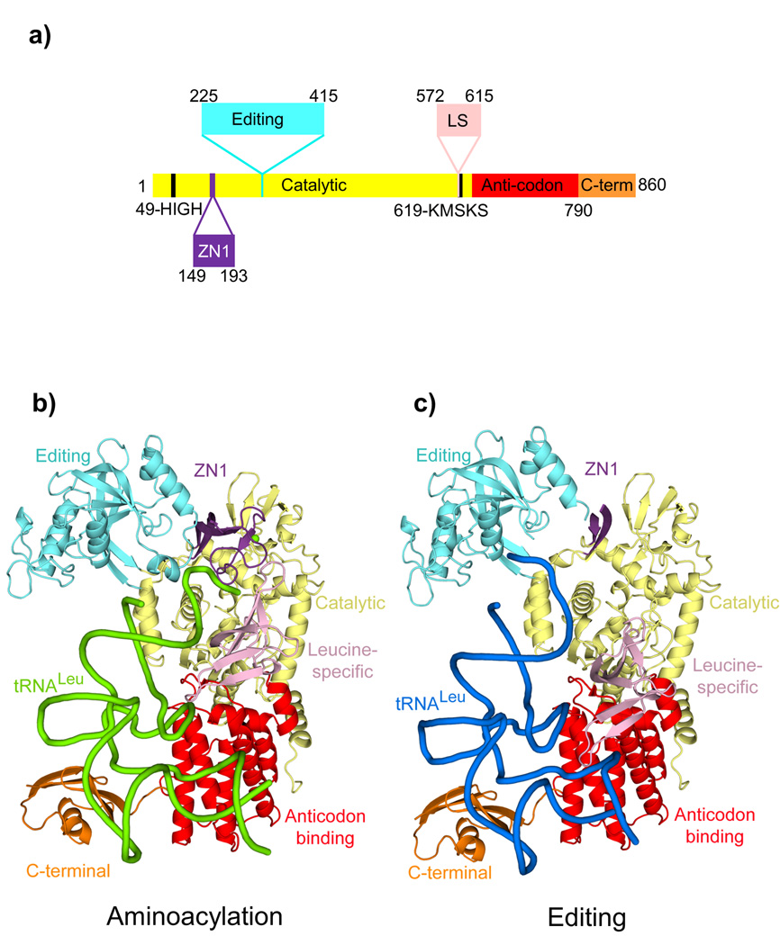
Figure 1. Structures of the E. coli LeuRS-tRNALeu complex in the aminoacylation and editing states.
a. The domain structure of LeuRSEC. Residue numbers indicate domain boundaries. The color code used throughout this paper for the various domains is catalytic (yellow), zinc (ZN1) (purple) with the zinc ion in green, editing (cyan), leucine-specific (pink), anticodon-binding (red) and C-terminal (orange).
b. Aminoacylation conformation with the tRNA in green.
c. Editing conformation with the tRNA in blue. In this state, the ZN1 domain is partially disordered.

