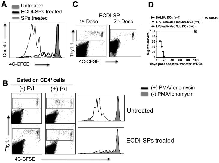
Figure 6. Donor ECDI-SPs infusions induce anergy in T cells with direct allo-antigen specificity.
4×10⁶ CFSE-labeled Thy1.1+CD4+ 4C T cells were adoptively transferred into Thy1.2+ recipients on day −8, followed by our standard tolerance induction with ECDI-SPs infusions (day −7, +1) and islet transplantation (day 0). 4C T cells were examined on day −4 (for A, B) and day +7 (for C). A, Histogram shows 4C T cell proliferation by CFSE dilution in mice: treated with donor ECDI-SPs (thick black line), with donor SPs not treated with ECDI (thin gray line), and untreated (shaded gray). B, T cells were isolated from the spleen of untreated mice (top panels) or mice treated with donor ECDI-SPs (bottom panels), and stimulated ex vivo with PMA and Ionomycin for 4 hours. Plots were gated on total CD4+ T cells. Dot plots show CSFE dilution of 4C cells before ((−)P/I) and after ((+)P/I) PMA/Ionomycin stimulation. Comparison of before and after PMA/Ionomycin stimulation is shown by histogram overlay. C, Further proliferation of the Thy1.1+ 4C T cells after receiving the 2nd dose of donor ECDI-SPs (day +1) was examined on day +7, and compared with that after the 1st dose of donor ECDI-SPs (examined on day −4). D, Long-term tolerized mice by ECDI-SPs infusions (>100 days graft survival post initial islet transplantation) were adoptively transferred with un-activated donor (BALB/c) BM-DCs, or LPS-activated donor (BALB/c) or third party (SJL) BM-DCs. Graft survival was monitored by blood glucose measurements. Data shown in A–C are representative of at least 3 individual experiments.

