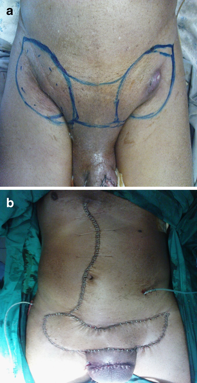
Fig. 3.
a Preoperative view of bilateral enlarged inguinal lymph nodes in a male patient following partial amputation in a case of carcinoma penis. Note the marking of the site of inguinal lymphadenectomy alongwith the involved overlying skin. b. Postoperative view of vertical rectus abdominis flap covering the bilateral inguinal defect after the flap has been rotated. Note prolene mesh has been used to cover the defect and the donor site has been closed primarily

