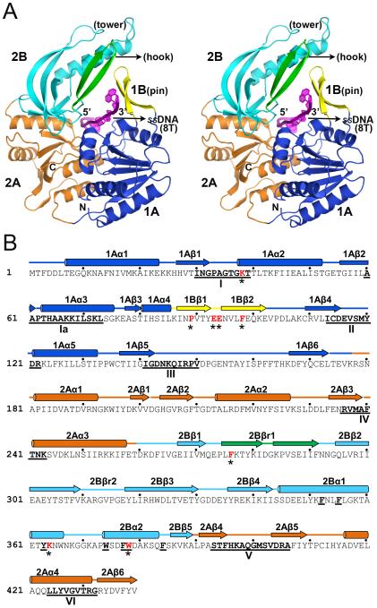
Figure 1. Structural overview of the Dda-ssDNA complex.
(A) Secondary structure representation of the complex showing the key structural and functional domains and motifs in stereo view. The 1A and 2A RecA-like domains are blue and orange, respectively, the SH3 domain (2B) including the tower is cyan, the β-ribbon pin (1B) is yellow, and the β-ribbon (hook) extension from 2B is green. The ssDNA is shown in magenta passing through the central arch. (B) The primary structure of Dda with the secondary structures labeled and colored as in (A). The helicase motifs are underlined in bold and labeled. Residues mutated in the study are shown in red with an asterisk. Key hydrophobic residues in the tower region (Figure 2) are bold and underlined.

