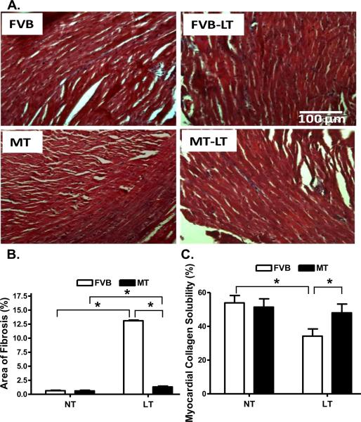
Fig. 5.
Histological analyses of myocardial fibrosis in FVB and metallothionein (MT) transgenic mice maintained at low temperature (LT, 4°C) for 3 months. A: Representative Masson Trichrome staining micrographs showing longitudinal sections of left ventricular myocardium (× 200); B: Quantitative analysis of fibrotic area (Masson trichrome stained area in light blue color normalized to the total myocardial area) from ~ 100 sections from 4 mice per group; and C: Myocardial collagen crosslinking from 6 mouse hearts per group. Mean ± SEM, * p < 0.05, NT: normal temperature.

