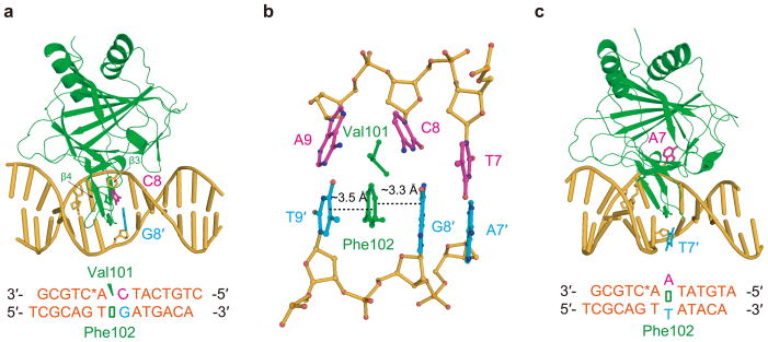
Figure 1.
Base pairs with different stability are discernible by ALKBH2. (a) Cartoon of the CG structure. (b) Local view showing the interrogation of the target C8:G8′ pair by ALKBH2, with residues Val101 and Phe102 highlighted. (c) Overall view of the AT structure. ALKBH2 is shown in green, DNA in yellow-orange, DNA bases from the upper strand in light magenta and those from the bottom strand in cyan.

