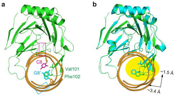
Figure 3.
ALKBH2 probes the stability of a base pair to detect DNA damages. (a) Side view of the CG structure. The approximate location of a flipped base is indicated with a magenta box and that of the orphaned base (which could have multiple conformations) is indicated with a dashed cyan box. (b) Overlay of the 1-meA structure (3BTY) and the CG structure. A clear shift of the hairpin loop is highlighted. The protein portion of 3BTY is shown in cyan and the DNA part is omitted for clarity purpose.

