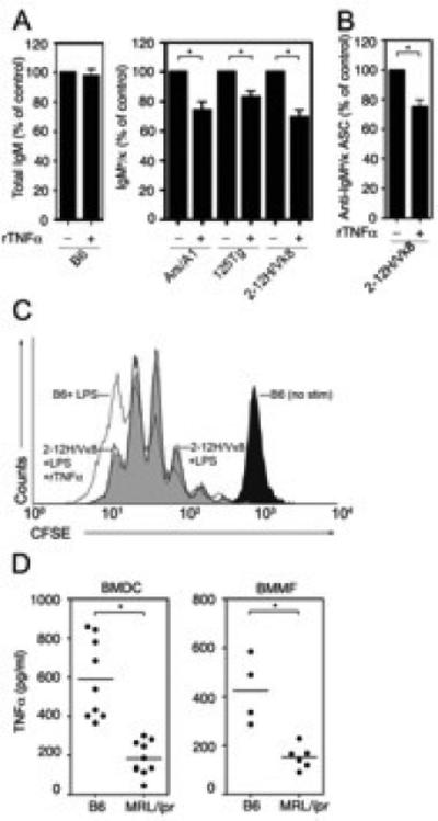
Figure 3. TNFα represses autoantibody secretion by reducing the number of ASCs.

(A) Purified B cells from the indicated mice were stimulated with LPS (30 μg/ml) and cultured with rTNFα (50 ng/ml). Total IgM (left) or IgMa/κ (right) was measured by ELISA from day four culture supernatant. LPS-stimulated B cells (100%) secreted 23-51μg/ml total IgM or 1-6 μg/ml IgMa/κ. ((B) Sm-specific (2-12H/Vκ8) B cells were stimulated with LPS (30 μg/ml) in the presence or absence of rTNFα (50 ng/ml) for 3 days. The frequency of ASCs was determined by ELISPOT. (C) CFSE loaded B cells from 2-12H/Vκ8 mice were incubated in the presence (black line) or absence (gray shade) of rTNFα (50 ng/ml). The proliferation indices were 5.6 and 5.5, respectively. The CFSE dilution of LPS stimulated is shown as a reference (D) BMDCs (left) or BMMF (right) from B6 or MRL/lpr were stimulated with LPS (30 μg/ml). TNFα was quantitated by ELISA from day four culture supernatants. Data represent at least three independent experiments. Error bars depict SEM. (* p≤0.05).
