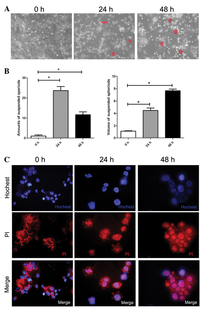
Figure 1.

Anoikis of suspended SMMC-7721 cells and formation of multi-cellular spheroids. (A) Morphology of SMMC-7721 cells in agar suspension culture at indicated time-points (magnification, ×100). (B) Characterization of the suspended spheroids during suspension culture of SMMC-7721 cells. The amount of spheroids (3 cells/spheroid) per focus was counted under a phase-contrast microscope. The volume of spheroids referred to the average number of cells per multi-cellular spheroids (magnification, ×100; n=8–10). Each value is the mean ± SD of at least triplicate determinations. (C) Double staining of Hoechst 33342 and PI SMMC-7721 cells after suspension at different time-points by immunofluorescence under a laser scanning microscope (magnification, ×400).
