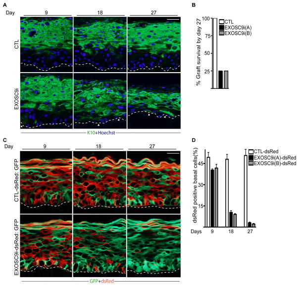
Figure 2. EXOSC9 loss results in premature differentiation and loss of self-renewal in human epidermal tissue.
(A) Keratinocytes expressing shRNAs for EXOSC9 (EXOSC9i) or control shRNA (CTL) were used to regenerate human epidermis on immune deficient mice. Experiments were performed using two separate shRNAs targeting different regions of EXOSC9i [EXOSC9i(A) and EXOSC9i(B)]. Tissues were harvested at 9, 18, and 27 days post grafting on mice. Keratin 10 (K10) staining shown in green marks differentiated epidermal layers. Hoechst staining in blue marks the nuclei. White arrowheads denote areas of ectopic basal layer differentiation. The dashed lines denote basement membrane zone (Scale bar=40μm; n=4 grafted mice per shRNA construct per timepoint). (B) Graft survival. Knockdown grafts that survived on mice by day 27. (C) Human epidermal progenitor competition assay. Keratinocytes were first transduced with a retrovirus to constitutively express dsRed. dsRed expressing cells were then knocked down for EXOSC9 (EXOSC9i) or control (CTL) and mixed at a 1:1 ratio with GFP expressing keratinocytes. The mixed cells were used to regenerate human epidermis on immune deficient mice. GFP expressing cells are shown in green while dsRed expressing cells are shown in red. Scale bar=40μ n=4 grafted mice per shRNA construct per timepoint. (D) Quantification of dsRed cells in the basal layer. Error bars=mean with SEM. See also Figure S1 and S2.

