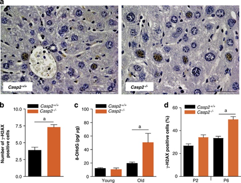
Figure 2.
Old Casp2−/− mice display increased DNA damage. (a) Immunohistochemical localization of γ-H2AX in aged mice liver ( × 40), scale bar=50 μm. (b) Quantitation of γ-H2AX stained cells in liver sections from old Casp2+/+ and Casp2−/− mice. Values are average number of γ-H2AX positive cells per field of view at × 40 magnification. (c) 8-OHdG levels in liver DNA from young and old Casp2+/+ and Casp2−/− animals (n=5 or 4) were determined by using an EIA kit (Cayman Chemicals). Values are mean±S.E.M. aP<0.05 represents comparison between the two groups. (d) Casp2−/− MEFs at early passage (P2) and late passage (P6) stained with γ-H2AX after menadione treatment and the frequency of γ-H2AX positive cells quantitated. Values are mean±S.E.M. aP<0.05 represents comparison between the groups

